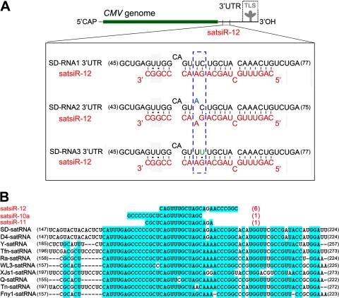
Fig. 2.
Imperfect complementarity between satsiR-12 and the 3′ UTR of SD-CMV RNA and alignment of representative CMV satRNA strains with satsiRNAs. (A) Sequence alignment among satsiR-12 and the SD-CMV RNA1, RNA2, and RNA3 3′ UTRs upstream of the TLS region. 5′CAP represents the 5′-terminal cap structure. The three mismatches between satsiR-12 and SD-CMV RNA1 and RNA3 and four mismatches between satsiR-12 and RNA2 are shown. The differences among the three target regions are indicated in the dashed box. (B) Alignment of representative CMV satRNA isolates/strains with satsiRNAs. Three cloned satsiRNAs (17) mapped to the conserved region (highlighted with a blue background) are shown. Red numbers in parentheses indicate the clone frequencies for each satsiRNA in this conserved region (17). Black numbers at the ends of the sequences indicate nucleotide positions.

