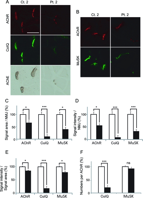
Figure 4. Passive transfer of muscle-specific receptor tyrosine kinase (MuSK)–immunoglobulin G (IgG) of control 2 and patient 2 to C57BL/6J mice.
(A, B) Quadriceps muscle sections of mice injected with IgG of control 2 or patient 2 are stained for acetylcholine receptor (AChR) by Alexa594-labeled α-bungarotoxin, collagen Q (ColQ) and MuSK by immunostaining, acetylcholinesterase (AChE) by cytochemical staining. Scale bar = 40 μm. Signal areas (C), intensities (D), and densities (intensity/area) (E) of the indicted molecules per neuromuscular junction (NMJ) are shown in mean and SEM. (F) Densities of ColQ and MuSK are normalized for the density of AChR to estimate the number of ColQ and MuSK per AChR. For AChR, ColQ, and MuSK, we analyzed 44 NMJs of control 2 and 23 NMJs of patient 2. For MuSK, we analyzed 82 NMJs of control 2 and 42 NMJs of patient 2. Areas and intensities are quantified by the BZ-9000 microscope (Keyence). Open and closed bars represent control 2 and patient 2, respectively. *p < 0.05, ***p < 0.001. NS = not significant.

