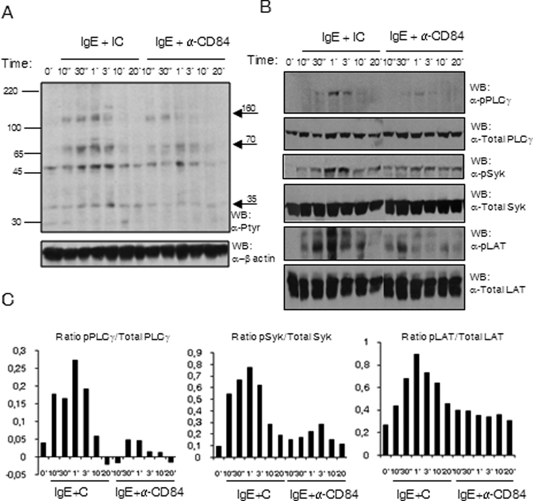
Figure 5. (A) FcεRI and CD84 co-engagement reduce the phosphorylation of specific proteins.

Phosphotyrosine western blot of LAD2 cell lysates upon biotinylated IgE and anti-CD84 or isotype control crosslinking with 250 ng/ml SA. The time of stimulation is indicated in the figure. Approximate molecular weight of differentially phosphorylated proteins are indicated by arrows on the right (B) SykY352, LATY191 and PLCγ1Y783 are less phosphorylated upon receptors co-ligation. Specific anti-phosphoresidue antibodies for pSykY352, pLATY191 and pPLCγ1Y783 were used to blot membrane with LAD2 cell lysates upon biotinylated IgE and anti-CD84 or isotype control crosslinking with 250 ng/ml SA. The time of stimulation is indicated in the figure. Membranes were reprobed with anti-total PLCγ1, anti-total Syk and anti-total LAT Abs to calculate phosphorylated/total protein ratios and to check loading levels per lane. Densitometric analysis and phosphorylated /total ratio for each molecule from 3 independent experiments is represented in the bar charts on the bottom (C).
