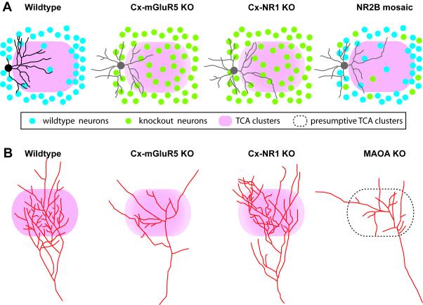
Figure 3.
Schematic diagram showing examples of reconstructed layer IV spiny stellate neurons, and single thalamocortical axons in wildtype, Cx-mGluR5 KO, Cx-NR1 KO and NR2B mosaic mutant mice. (A) In wildtype mice, layer IV spiny stellate neurons show polarized dendritic morphology with the majority of their dendrites projecting toward the barrel hollow (pink). For simplicity, only spiny stellate neurons are depicted here. In both Cx-mGluR5 and Cx-NR1 KO mice, layer IV neurons are evenly distributed in the barrel field, and have symmetric dendritic morphology (non-polarized pattern, depicted in grey). In NR2B mosaic KO mice, mutant layer IV neurons located in the barrel walls show non-polarized dendritic pattern. (B) Examples of reconstructed single TCAs. In wildtype mice, the axon arbor in layer IV develops from a single axon, forming numerous axon collaterals, with a dominant orientation of the branches toward the barrel center, individual TCAs form highly branched and densely clustered arbors corresponding to the mapped facial whisker. In Cx-mGluR5 KO mice and MAOA KO mice, the complexity of TCA arbors is much reduced, and collaterals grow in divergent directions instead of forming a narrow cluster. In Cx-NR1 KO mice, TCAs have exuberant branches. Drawings were based on data shown in Datwani et al. (2002), Rebsam et al. (2002), Lee et al. (2005), Espinosa et al. (2009), and Ballester-Rosado et al. (2010).

