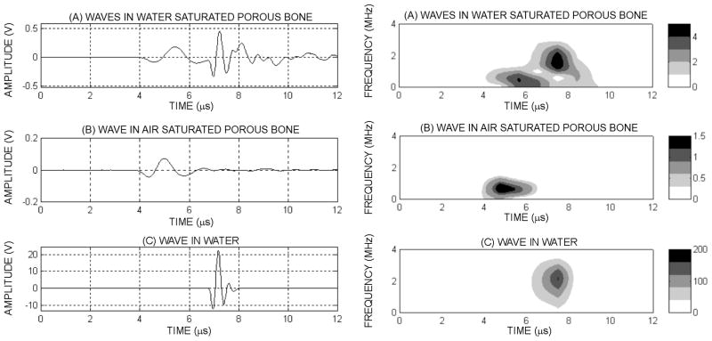
Figure 3.
Ultrasound wave after propagation through a fluid saturated human cancellous bone sample (a) signal propagated through the same human sample after the water was removed from the pores (b), and detected pulse after propagating in water on a distance identical to the sample’s size (c). Corresponding spectrograms of a human signal showing the two waves having different frequency compounds and time localization (d), when the fluid is removed from the pores (e) and when the porous sample is removed and the wave propagates in the fluid only (f). The color bar indicates the respective power spectra density value (Vrms2)

