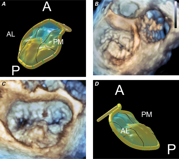
Fig. 1 A) Topographic model of P2 and P3 (posterior leaflet prolapse). Posteromedial (PM)-to-anterolateral (AL) projection shows the right fibrous trigone (PM) and left fibrous trigone (AL) in systole. B) Three-dimensional transesophageal echocardiographic reconstruction in the same projection shows the aortic valve just beginning to open after the posterior mitral leaflet prolapses in systole. The tricuspid valve is on the right. C) Three-dimensional transesophageal echocardiographic reconstruction (left atrial frontal view) shows a nearly 3-cm prolapsing segment of P2. D) Topographic model of P2 and P3 (posterior leaflet prolapse): AL-to-PM projection is shown in systole.
A = anterior; P = posterior

