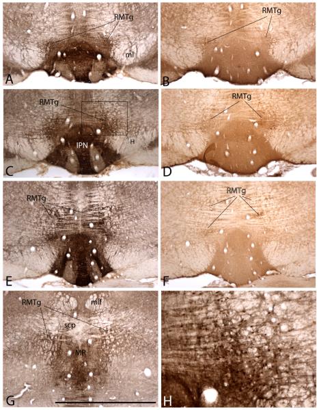
Figure 2.
Photomicrographs of sequentially more caudal sections showing the RMTg in preparations processed to exhibit the μ-opioid receptor (A, C, E, G and H) and somatostatin (B, D and F) immunoreactivities at levels comparable to those shown in Figure 1A, B, C and E. H is an enlargement of the box shown in C and illustrates Fos immunoreactive nuclei (brown dots), which are concentrated in the RMTg in rats, which, like the one from which these preparations were made, received an injection of psychostimulant drug prior to being sacrificed. Abbreviations: IPN – interpeduncular nucleus; ml – medial lemniscus; mlf – medial longitudinal fasciculus; MR – median raphe nucleus; RMTg – rostromedial tegmental nucleus; scp – superior cerebellar peduncle. Scale bar: 1 mm for A, -G; 250 μm for G.

