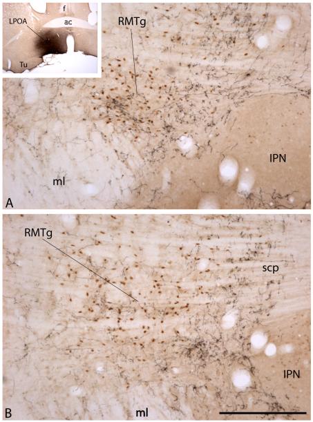
Figure 4.
Photomicrographs showing the RMTg at a rostral (A) and more caudal (B) level. Note the position of the collection of Fos–immunoreactive neuronal nuclei (brown dots) representing the RMTg, which occupy the angle between the medial lemniscus (ml) and interpeduncular nucleus (IPN) in A and lie above and lateral to the IPN in the more caudal section shown in (B). Levels shown in (A) and (B) are comparable to those in (A) and (C) in Figure 2. The RMTg is Fos immunoreactive in this preparation due to the fact that the rat from which the preparation was made was injected with the psychostimulant drug, methamphetamine (10 mg/kg), prior to being sacrificed. Ten days before sacrifice, the rat received an injection of anterogradely transported Phaseolus vulgaris-leucoagglutinin (PHA-L) into the lateral preoptic area (LPOA), resulting in anterograde labeling of a dense plexus of axons in and around the RMTg (black labeling). Note the horizontal banding pattern of the crossing fibers of the superior cerebellar peduncle (scp in B). Scale bar: 200 μm. Additional abbreviations: ac – anterior commissure; f – fornix; Tu – olfactory tubercle.

