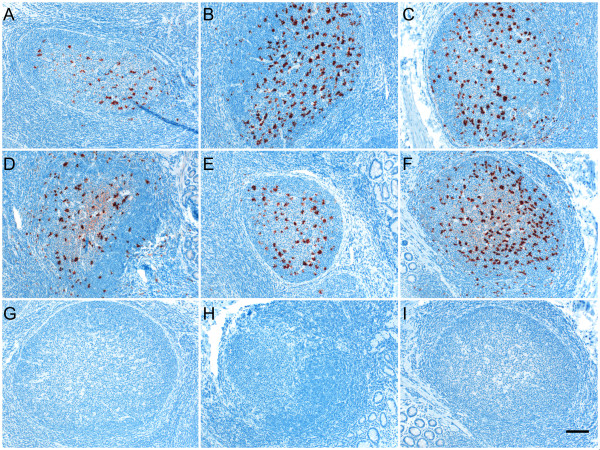
Figure 1.
Immunolabeling of PrPSc in the rectoanal mucosa-associated lymphoid tissues of recipient sheep transfused with different blood components. PrPSc immunolabeling (dark red) was visible in the RAMALT follicles of recipient sheep receiving either whole blood (A), buffy coat (B), PBMCs (C), CD72+ B lymphocytes (D), CD21+ B lymphocytes (E) or platelet-rich plasma (F) fractions but not after receiving a platelet-poor plasma (G) fraction when labeled with anti-prion mAbs. PrPSc immunolabeling was not observed in RAMALT follicles of scrapie negative (H) sheep or when using an isotype-matched control mAb (I; same tissue block shown in F). Scale bar = 50 μm.

