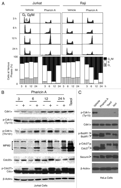
Figure 3.
Pharicin A-induced mitotic arrest is correlated with the activation of Cdk1. (A) Jurkat and Raji cells were treated with pharicin A (2 µM) for the indicated times for various times as indicated. DNA content of the treated cells was analyzed by flow cytometry. (B) Jurkat cells were treated with pharicin A (2 µM) for various times were collected and approximately equal amounts of cell lysates were blotted for Cdk1, p-Cdk1(Tyr 15), p-Cdk1(Thr 161), MPM-2, Cdc20, Cdc27 and β-actin. Cells treated with paclitaxel for 24 h were lysed and equal amounts of cell lysates were used as control. (C) HeLa cells were treated with pharcin A, paclitaxel (Taxol) or nocodazole (Noc) or vehicle for 24 h were collected and lysed. Equal amounts of cell lysates were blotted for Cdk1, p-Cdk1(Tyr15), BubR1, Cdc27, securin and β-actin.

