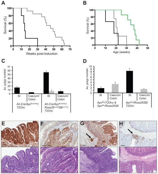
Figure 1. SB transposon mobilization increases morbidity and tumor burden of Apc-deficient mice.
(a) Kaplan-Meier survival curve comparing Apcfloxed experimental (Ah-Cre/Apcfloxed/wt/Rosa26Lox66SBLox71/T2Onc) (black line) and control mice (Ah-Cre/Apcfloxed/wt/T2Onc and Ah-Cre/Apcfloxed/wt/Rosa26Lox66SBLox71) (grey line) (log-rank P<0.001). (b) Kaplan-Meier survival curve comparing ApcMin experimental (ApcMin/RosaSBase/T2Onc) (black line), ApcMin control mice (ApcMin/RosaSBase and ApcMin/T2Onc) (grey line) and RosaSBase/T2Onc control mice (green line) (log-rank P<0.0057). (c) Average polyp size and number in Apcfloxed experimental mice (n=3), and matched littermate Apcfloxed control mice (n=3) at 18 weeks post-induction. All polyps visible under the dissecting microscope at 6x magnification were counted and grouped into those that were non-excisable (black bars) (<3mm) and those that could be excised for further analysis (>3mm) (grey bars). Average polyp number for small intestine and caecum/colon is shown. (d) Average number of excisable polyps (>3mm) from small intestine, caecum and colon of ApcMin experimental mice (n=10) and ApcMin control mice (n=7) at 10-22 and 17-30 weeks of age respectively. β-catenin immunohistochemistry (upper panel of each) and hematoxylin and eosin staining (lower panel of each) of representative tissue from Apcfloxed mice showing (e) a tubulovillous adenoma at 18 weeks post-induction, (f) an adenoma at 28 weeks post-induction, (g) an adenoma at 17 weeks post-induction with the arrow indicating the region of adenocarcinoma in the submucosa. (h) Lysozyme staining of representative tissue from an Apcfloxed/wt mouse at 22 weeks post-induction, with the arrow indicating Paneth cell differentiation. All scale bars = 100μm.

