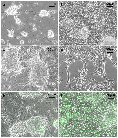
Figure 1. Neural cultures plated onto PLL- or AK-cyclo[RGDfC]- coated surfaces.
Phase-contrast view of primary cultures of fetal (E14.5) mouse forebrain cells on AK-cyclo[RGDfC]-coated surface, on the 2nd (a) and 6th (b) days after plating, and on poly-L-lysine (c) coated surface on the 6th day after plating. On AK-cyclo[RGDfC] morphologically homogeneous cultures of radial glia-like cells developed after the first passage (d). In primary cultures prepared from the forebrain of hGFAP-GFP mouse embryos (E14.5), GFP-expressing cells colonized the AK-cyclo[RGDfC] surface (f), while stayed inside the aggregates on PLL (e) (6th day after plating).

