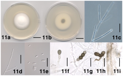
Figure 11. Morphological features of Verticillium nubilum.
11. Colony of strain P742 after 13 days on PDA, frontal view. 1b. Colony of strain PD742 after 13 days on PDA, reverse view. 11c. Conidiophore of strain PD621 after 17 days on WA-p. 11d. Apical phialide of strain PD621 after 17 days on WA-p. 11e. Conidia of strain PD621 after 17 days on WA-p. 11f. Solitary chlamydospore of strain PD742 after 17 days on WA-p. 11g. Linear chain of chlamydospores of strain PD742 after 17 days on WA-p. 11h. Angular chain of chlamydospores of strain PD621 after 25 days on PDA. 11i. Brown-pigmented hyphae of strain PD621 after 25 days on PDA. Scale bar: 11a, 11b = 1 cm; 11c–11i = 20 µm; Imaging method: 11a, 11b, = DS; 11c = PC; 11d, 11e = DIC; 11f–11i = BF.

