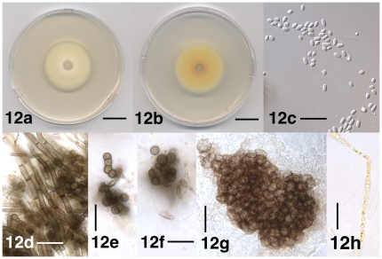
Figure 12. Select morphological features of Verticillium tricorpus.
12a. Colony of strain P685 after 10 days on PDA, frontal view. 12b. Colony of strain PD685 after 10 days on PDA, reverse view. 12c. Conidia of strain PD685 after 38 days on PDA. 12d. Resting mycelium of strain PD685 after 38 days on PDA. 12e. Chain of chlamydospores and microsclerotium of strain PD685 after 38 days on PDA. 12f. Microsclerotium of strain PD685 after 38 days on PDA. 12g. Microsclerotium of lectotype specimen IMI 51602. 12h. Yellow-pigmented hypha of strain PD685 after 38 days on PDA. Scale bar: 12a, 12b = 1 cm; 12c–12h = 20 µm; Imaging method: 12a, 12b = DS; 12c = DIC; 12d–12h = BF.

