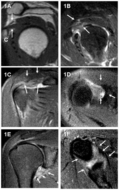
Figure 1. A 56-year-old female patient with clinical evidence of a right frozen shoulder.
Sagittal oblique T1-weighted image (TR/TE = 550 ms/15 ms) (1A) shows thickened CHL (arrows). C = inferior margin for the coracoid process. Sagittal oblique (1B), oblique coronal (1C), and transverse (1D) fat-suppressed, proton density weighted, spin-echo image (TR/TE = 3000 ms/34 ms) show high-signal intensity soft tissue in the rotator cuff interval for the same patient (arrows). Coronal oblique (1E) and transverse (1F) fat-suppressed, proton density, weighted spin-echo image (TR/TE = 3000 ms/34 ms) demonstrate a thickened inferior glenohumeral ligament (axillary recess) for the same patient (arrows).

