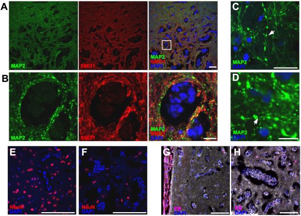
Figure 5. Neuronal injury and loss in co-opted 231BR brain metastases.
MAP2 staining displayed extensive beading in dendrites in lesions (A & B) indicative of acute injury, whereas staining for the phosphorylated form of neurofilaments (SMI31) did not show a similar pattern. Panel B is an enlargement of panal A. At high magnification (C & D) the beading phenomenon appeared as focal enlargements on continuous dendrites (arrows). Compared to the periphery of the lesion (E), the number of neuronal cell bodies was decreased in the metastatic core (F). Solid metastases in the ventricles (G), but not co-opted cells in the brain (G & H) incorporated intraveneously injected Evan’s blue dye. Scale bars indicate 100 μm in A, E, F, G, & H, and 20 μm in B, C, & D.

