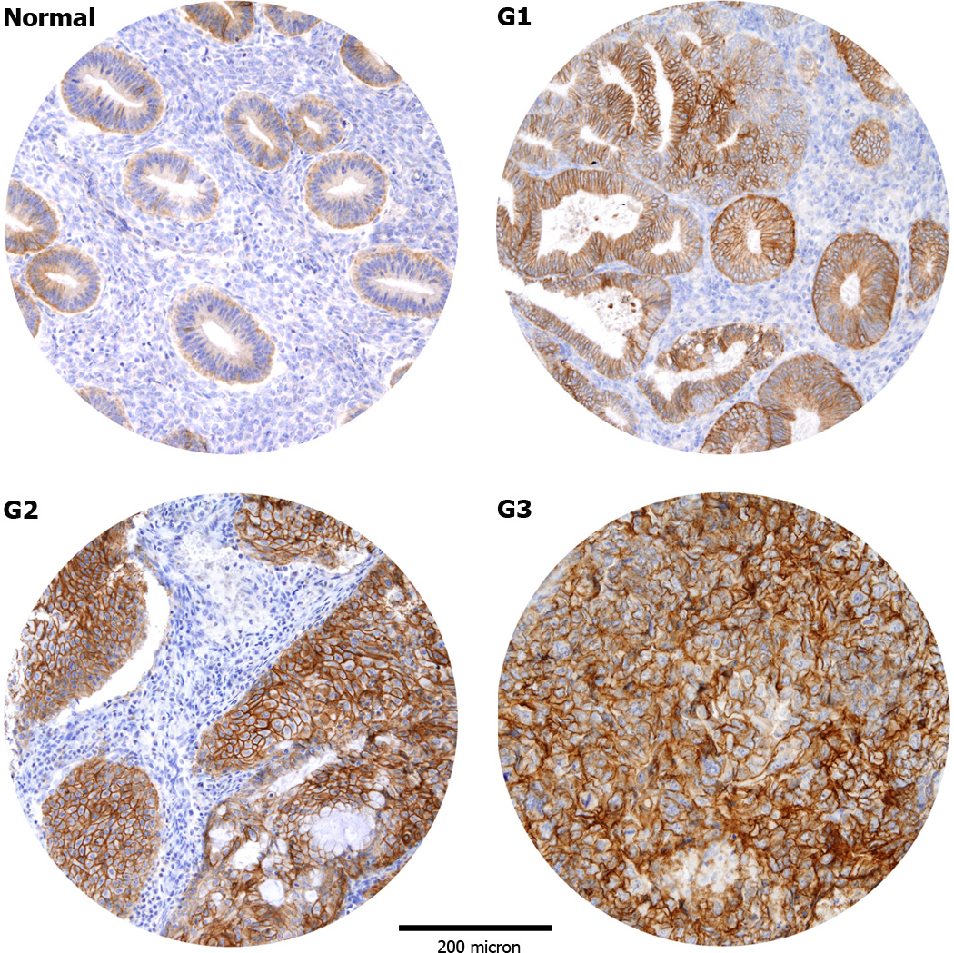
Figure 1.
Representative immunohistochemical staining for Trop-2 in tissue microarrays of normal endometrial controls (NEC) and endometrial carcinomas (EC). NEC tissues show predominantly a weak immunoreactivity for Trop-2 (A). Positive Trop-2 membrane staining (B–D): grade 1 (B), grade 2 (C), grade 3 (D) endometrioid ECs. (Original magnification X100). The figure were taken at the same magnification and the periphery of the core was resized using Adobe photoshop, with similar reduction of the total core area (>5%).

