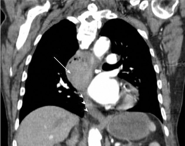
Figure 1.

Coronal reformat of a contrast-enhanced chest computed tomography demonstrates a solid enhancing mass with soft tissue attenuation in the middle third of the esophagus (arrow) causing obstruction and proximal dilatation. The distal third of the esophagus (arrowheads) is unremarkable.
