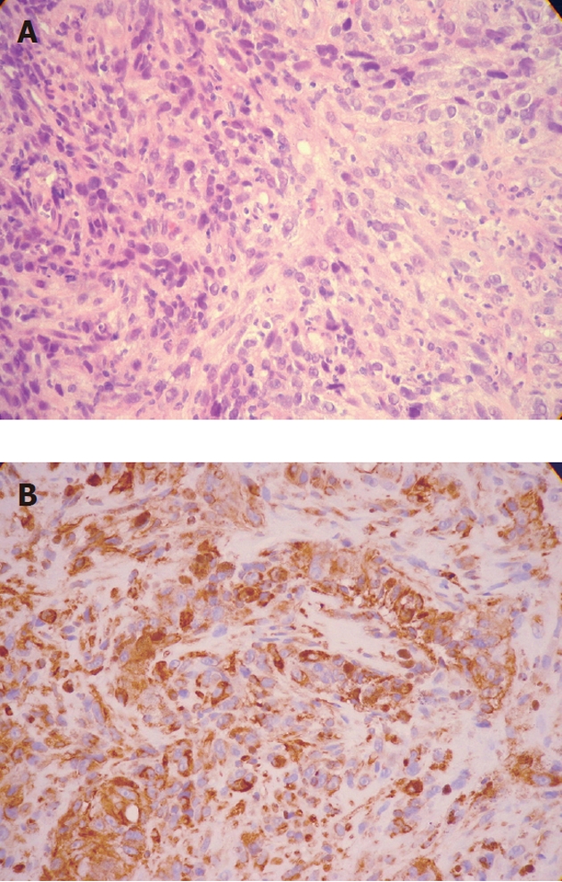
Figure 3.

Histopathological findings. A: Hematoxylin and eosin staining discloses crowded rounded or elongated cells within distinct cytoplasm and hyperchromatic nuclei (magnification, x 100); B: Immunohistochemical study for HMB45 (anti-melanoma protein mAb). The positive reaction (brown cytoplasmic reaction) supports the diagnosis of malignant melanoma (magnification, x 100).
