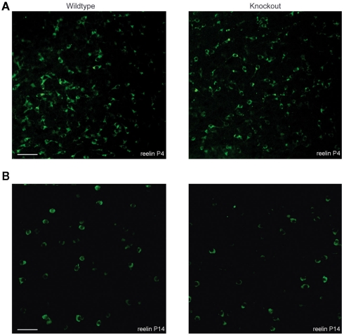
Figure 6.
Tangential sections showing the distribution of reelin-positive Cajal–Retzius cells in the somatosensory cortex of both wildtype and 5-HT3A receptor knockout mice. Typical examples of reelin-positive Cajal–Retzius cells in P4 (A) and P14 (B) tangential sections of the somatosensory cortex of both wildtype and 5-HT3A receptor KO mice. Scale bar 50 μm.

