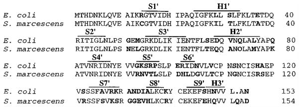
Figure 2.
Comparison of the amino acid sequence of the regulatory polypeptides between Ec and Sm ATCase. The standard single-letter abbreviations from the amino acids are used. The presence of a “.” indicates a shift in sequence to optimize the alignments. The residues in bold are those which differ between the Ec and Sm ATCases. The lines above the sequence correspond to the secondary structural elements as determined for the Ec enzyme with H representing α-helices and S representing β-strands. Each structural element is numbered sequentially with the “′” indicating its location in the regulatory polypeptide. The S5′ β-strand is comprised in its entirety by the altered region (r93–r97).

