Fig. 2.
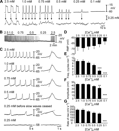
Oviduct slow wave generation is dependent on [Ca2+]o. A: simultaneous recordings of slow waves (top) and myosalpinx contractions (bottom) made during perfusion with 2.5–0 mM [Ca2+]o Krebs-Ringer bicarbonate buffer (KRB). Slow waves and contractions were coupled on a 1:1 basis (dashed arrows) and as [Ca2+]o was reduced, slow wave and contraction amplitude and frequency decreased until they were both abolished at concentrations lower than 0.25 mM [Ca2+]o. B: complete intracellular recording as [Ca2+]o was reduced from 2.5–0.25 mM when slow wave activity ceased. C: expanded time scales show the gradual reduction in frequency and rate of rise of slow waves (indicated by dashed lines on the expanded slow waves) as [Ca2+]o was reduced. D–G: summarized results of 7 experiments examining the effects of reduced [Ca2+]o on slow wave parameters. Resting membrane potential (RMP, D), slow wave frequency (E), slow wave amplitude (F), and maximum rate of rise of the upstroke (G) are plotted as a function of [Ca2+]o. Statistically significant changes in slow wave parameters are indicated: *P < 0.05, **P < 0.01, ***P < 0.001 (one-way ANOVA with a post hoc Tukey test). NB 0 mM [Ca2+]o refers to nominally free calcium. Negligible amounts of Ca2+ may have been present in salts, other than CaCl2, present in KRB.

