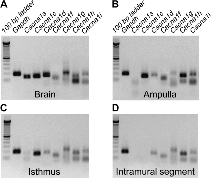
Fig. 3.
Expression of Ca2+ channel transcripts in the oviduct myosalpinx. Panels show RT-PCR gel analysis of Canca1s, Cacna1c, Cacnc1d, and Cacna1f encoding L-type (Cav1 subfamily) and Cacna1g, Cacna1h, and Cacna1i encoding T-type (Cav3 subfamily) expression in the ampulla (B), isthmus (C) and intramural (D) regions of mouse oviduct. Brain was used as a positive control tissue for each channel expression (A). The amplification of GAPDH was used as a control for cDNA integrity. The sizes of RT-PCR amplicons are indicated in Table 1.

