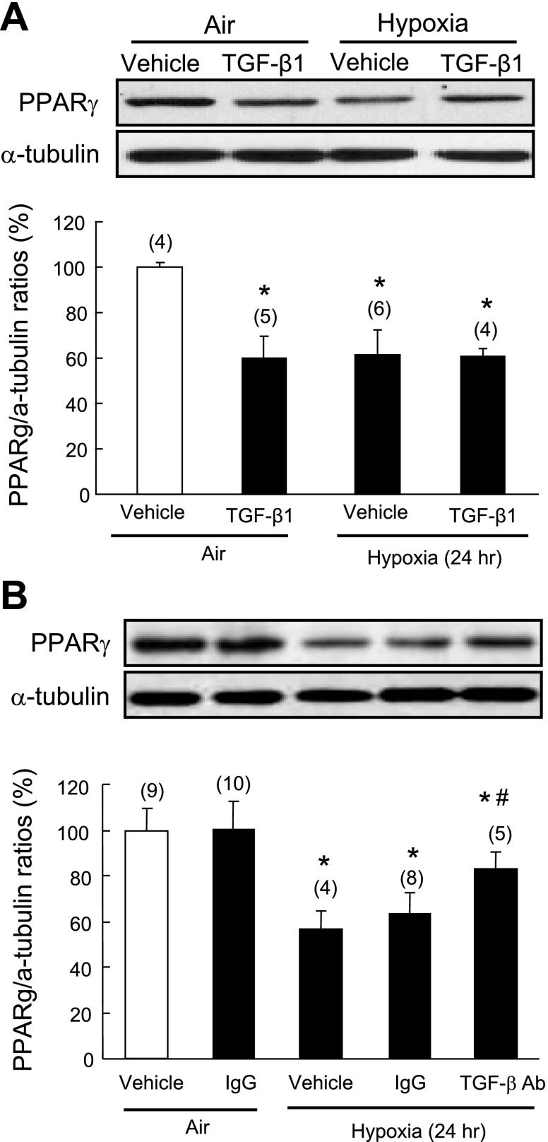
Fig. 4.
Effect of TGF-β1 on hypoxia-induced PPAR-γ downregulation in isolated pulmonary artery small muscle cells (PASMCs). A: quiescent rat PASMCs were pretreated with TGF-β1 (2 ng/ml) for 1 h and then exposed to normoxia or hypoxia (1% O2) for an additional 24 h. B: quiescent rat PASMCs were pretreated with TGF-β neutralizing antibody (50 μg/ml) or normal IgG for 1 h before normoxic (air) or hypoxic exposure for 24 h. Cellular PPAR-γ expression was determined by Western blot analysis and normalized using α-tubulin. Results are means ± SE; n = number of wells. *P < 0.05 compared with vehicle in air control group; #P < 0.05 compared with vehicle in hypoxia group.

