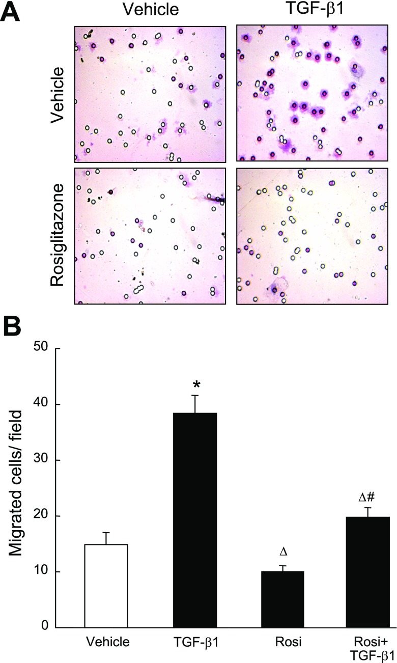
Fig. 8.
Effect of Rosi pretreatment on TGF-β1-induced migration of PASMCs. Quiescent PASMCs (0.5 × 10⁵) were seeded in Falcon cell culture inserts (8.0-μm pore size), pretreated with Rosi (0.1 μM) or vehicle for 3 h, then stimulated with TGF-β1 (2 ng/ml) for an additional 12 h. The cells adherent to the outer surface of the lower side of the membrane were counted. A: micrographs (×400) of the hematoxylin-stained cells that migrated through the pores. B: bar graphs summarize the number of PASMCs adherent to the lower side of the chamber. Results are means ± SE, n = 15 wells/group. *P < 0.05 compared with vehicle group; ΔP < 0.05 compared with TGF-β1 alone group; #P < 0.05 compared with respective Rosi alone group.

