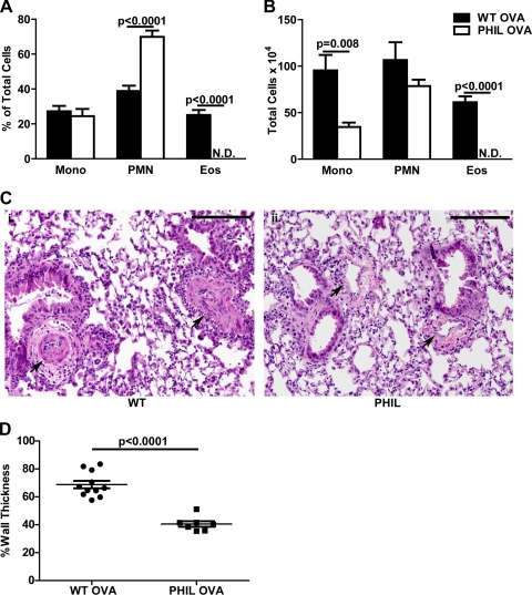
Fig. 2.
PHIL mice have attenuated pulmonary vascular remodeling. A and B: percentage and number of mononuclear cells, neutrophils, and eosinophils in the BAL of wild-type (WT) and PHIL mice following OVA immunization and challenge (n = 8–11 mice/group). C: representative hematoxylin- and eosin-stained lung sections of WT (i) and PHIL (ii) mice (×100 magnification). Pulmonary arteries are indicated with black arrows. Black bars are 100 μm (representative images from n = 8–11 mice/group). D: vessel wall thickness (% of total) in preacinar blood vessels in lung sections from WT and PHIL mice after OVA immunization and challenge (n = 8–11 mice/group). N.D., not detected.

