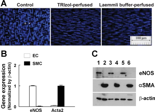
Fig. 4.
A: DAPI-stained vessels in control conditions (left), and after perfused with TRIzol (middle) or Laemmli buffer (right). Endothelial (EC) and smooth muscle cells (SMC) are aligned vertically (arrowhead) and horizontally (arrow), respectively. B and C: expression of endothelial nitric oxide synthase (eNOS) and α-smooth muscle actin (αSMA) mRNA by RT-PCR (n = 5) and protein by Western blotting (n = 3/3) in the endothelial lysates (EC) collected from TRIzol- or Laemmli buffer-perfused vessels, and SMCs (the remaining portions of vessels after collection of endothelial lysates). C: lanes 1, 3, and 5 were loaded with endothelial protein, and lanes 2, 4, and 6 were loaded with protein of SMC.

