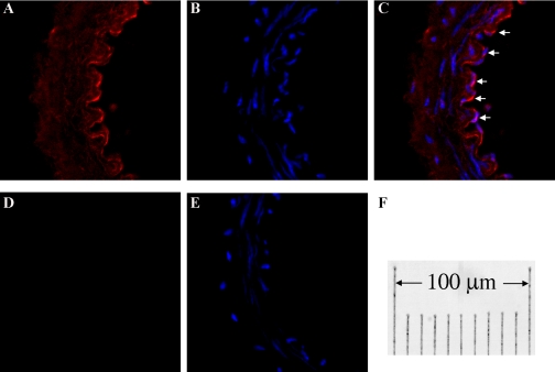
Fig. 6.
Sections of mesenteric arteries (endothelial layer faces to the right) of female l-NAME-treated rats were stained for CYP2C7 and nuclei with Cy3 (red) and DAPI (blue), respectively. A and B: section stained for CYP2C7 and nuclei, both of which are merged shown in C, indicating a prominent staining for CYP2C7 in ECs (arrows). D: section for negative control staining, which was additionally stained with DAPI shown in E. F: the final magnification.

