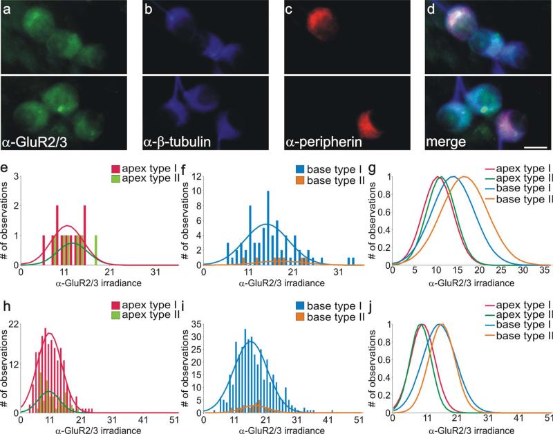
Figure 1.
Type I and type II spiral ganglion neurons in vitro were immunolabeled similarly with anti-GluR2/3 antibody. a: Two different fields from the same culture dish show that basal type I and type II spiral ganglion neurons in vitro were comparably stained with anti-GluR2/3 antibody (green). b: Both types of spiral ganglion neurons were also stained similarly with anti-β-tubulin antibody (blue). c: Anti-peripherin antibody labeling of presumptive type II neurons (red). d: Merged image of neurons in a and b show that, although clusters of neurons contained both putative type I and type II neurons, there were no significant differences in their staining patterns with anti-GluR2/3 and anti-β-tubulin antibodies. e,f: Frequency histograms of anti-GluR2/3 antibody irradiance measurements from a single experiment; apical type I and type II neurons and basal type I and type II neurons were all well fitted with single Gaussians. g: Normalized Gaussian fits for histograms in e and f show that average irradiance levels of both basal type I and type II neurons are greater than those obtained from both classes of apical neuron. h,i: Frequency histograms composed of anti-GluR2/3 antibody irradiance measurements from six experiments; apical type I and type II neurons and basal type I and type II neurons were well fitted with single Gaussians. j: Normalized Gaussian fits for histograms shown in h and i. Type I and type II distributions were essentially overlapping from each area. As in the single experiment, irradiance levels were consistently highest for both classes of basal neuron. For a magenta-green version see Supporting Information Figure 1. Scale bar = 10 μm.

