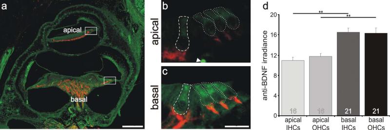
Figure 5.
Inner and outer hair cells located in the basal cochlea had a higher level of anti-BDNF antibody labeling than those located in the apical cochlea. a: Low-magnification view of a postnatal tissue section labeled with anti-BDNF (green) and anti-β-tubulin (red) antibodies. b,c: High-magnification merged images of sensory hair cells outlined in a (white boxes). Regions of measurement are demarcated with a dashed line for inner hair cells and dotted lines for outer hair cells. d: Averages of 15 and 20 measurements (apex and base, respectively) taken from 10 separate sections from three different preparations with two anti-BDNF antibodies showed that irradiance was significantly different between the hair cells from the apical and basal cochlear. Significance of the paired Student's t-test, **P < 0.01. For a magenta-green version see Supporting Information Figure 3. Scale bars = 120 μm in a; 15 μm in c (applies to b,c).

