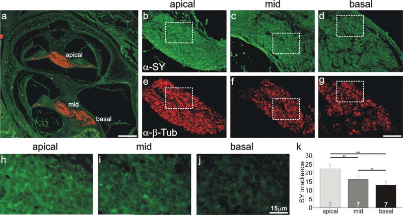
Figure 8.
Anti-synaptophysin antibody labeling of spiral ganglion neurons was systematically graded along the tonotopic axis. a: Low-magnification view of a postnatal cochlear section labeled with anti-synaptophysin (green) and anti-β-tubulin (red) indicating the location of spiral ganglion neurons in different cochlear turns. b–d: Anti-synaptophysin (green) antibody labeling of spiral ganglion neurons was highest in apical neurons (b), intermediate in a midcochlear region (c), and lowest in the base (d). e–g: Spiral ganglion neurons shown from b–d, respectively, were uniformly labeled with anti-β-tubulin (red). h–j: High-magnification view of neurons outlined with white boxes shown in b–d. k: Average values from seven experiments confirmed that anti-synaptophysin irradiance levels were highest in apical neurons and decreased systematically in neurons innervating more basal cochlear regions. Significance of the paired Student's t-test, *P < 0.05, **P < 0.01. For a magenta-green version see Supporting Information Figure 6. Scale bars = 150 μm in a; 40 μm in g (applies to b–g); 15 μm in j (applies to h–j).

