Figure 2.

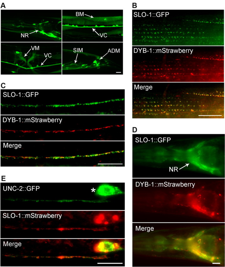
DYB-1 and SLO-1 were coexpressed and colocalized in neurons and muscle cells. A, Expression of GFP under the control of Pdyb-1 resulted in GFP epifluorescence in many neurons in the nerve ring (NR), ventral cord (VC), and tail (not labeled), as well as several types of muscles, including body-wall muscle (BM), vulval muscle (VM), stomatointestinal muscle (SIM), and anal depressor muscle (ADM). B–D, SLO-1::GFP and DYB-1::mStrawberry colocalized in body-wall muscle cells (B), the dorsal nerve cord (C), and the nerve ring (D) when they were expressed under the control of their native promoters. E, SLO-1::mStrawberry and UNC-2::GFP, which were expressed in GABAergic neurons, colocalized in the ventral nerve cord. * indicates the soma of a GABAergic motoneuron. Scale bars, 10 μm.
