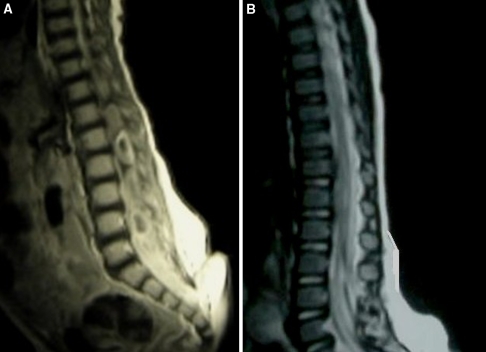
Fig. 3.
a Sagittal MRI scan T1 W scan with contrast of the thoraco-lumber spine showing intramedullary hypointense multicystic lesion extending from L1 to L5 with marked enhancement and spinal cord enlargement. Note again the dimple and dermal sinus tract as it attached the skin to the thecal sac. b Postoperative MRI scan revealing a significant resolution of the abscess

