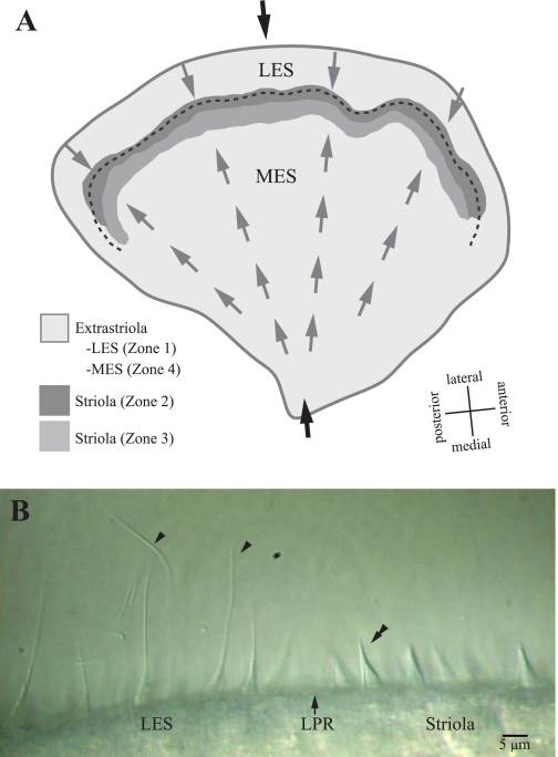
Fig. 1.
Turtle utricle. A: the macula is divided into 4 zones. Zone 1 corresponds to the lateral extrastriola (LES). Zones 2 and 3 form the striola: zone 2 (dark gray shading) is a 30- to 40-μm-wide band of type II hair cells with distinctive bundles; zone 3 (medium gray shading) is a 50- to 75-μm-wide band of type I hair cells. Zone 4 corresponds to the medial extrastriola (MES). The juxtastriola (not labeled) is an ∼100-μm-wide band just medial to zone 3. Hair bundle dimensions change gradually across zone 3 and the juxtastriola, from zone 2-like to zone 4 (MES)-like. The shapes and relative heights of bundles in different zones are summarized in Fig. 9 of Xue and Peterson (2006). Gray arrows indicate hair bundle polarity (axis of maximal sensitivity), and the dashed line indicates the line of polarity reversal (LPR). This LPR runs through zone 2, close to its lateral margin. Black arrows indicate the medial-to-lateral transect along which we folded the utricle to make stiffness measurements; it includes all macular subdivisions. B: hair bundles at the LPR. This differential interference contrast image shows the morphology of hair bundles immediately lateral (toward left in image) and medial (toward right) to the LPR (arrow). The LPR lies near the lateral edge of the striola and defines the line about which the excitatory directions of the hair bundles reverse orientation. Arrowheads mark 2 of the long kinocilia coupled to short stereocilia that are characteristic of LES hair bundles. The double arrowhead indicates a typical zone 2 hair bundle with a short kinocilium approximately as tall as the tallest stereocilia (KS ratio ∼1). Image contrast adjusted with ImageJ software.

