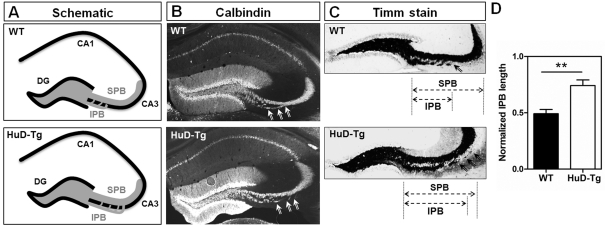
Figure 6. Increased length of the infrapyramidal mossy fibre bundle in HuD-Tg mice.
(A) Shows the distribution of IPB and SPB bundles in WT and HuD transgenic mice. Mossy fibres were stained using either calbindin antibodies (B) or Timm's stain (C). The IPB length was calculated from the hilus to where the fibres cross the pyramidal cell layer (arrows), and the length of the SPB was measured from the hilus to the apex of the curvature of area CA3 (C). The length of the IPB was normalized to that of the SPB in the same section (D). Scale bar = 300 μm. **P<0.01.

