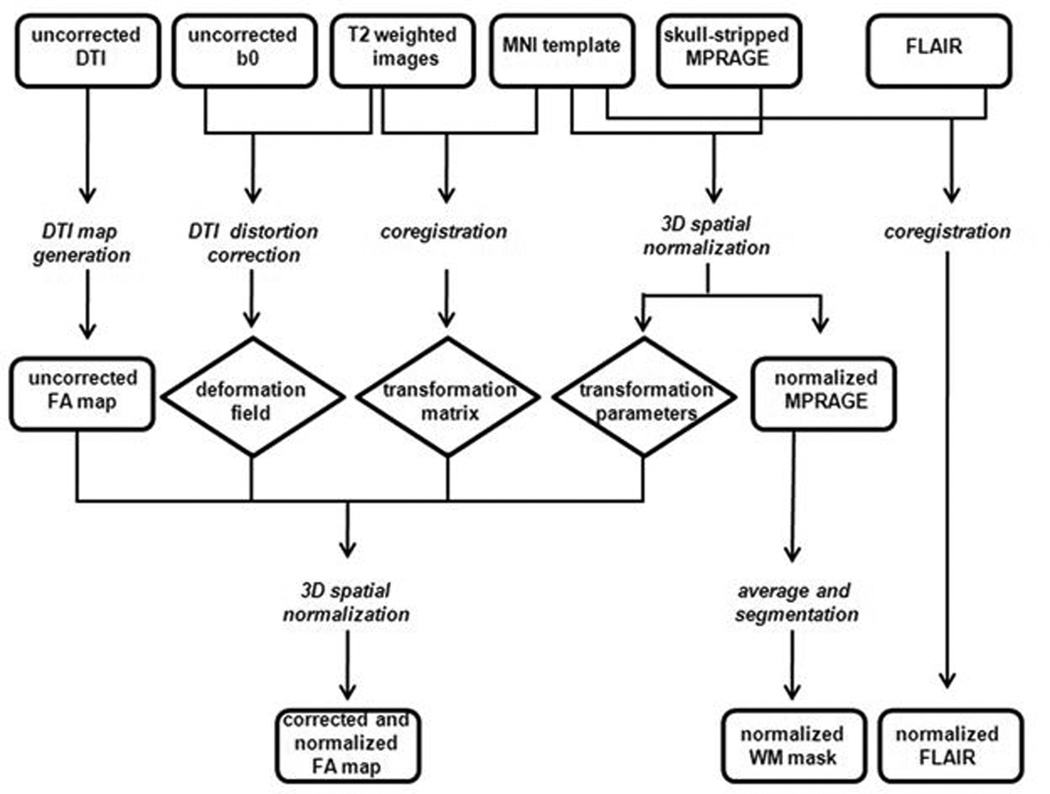
Figure 1. Generation of images for voxel wise analysis.
This is a pictorial depiction of how the originally acquired MR images were processed into a format where voxel wise analysis was possible. Rectangles represent images, italicized words represent procedures and diamonds represent the output from the procedures. Diffusion tensor imaging (DTI); Montreal Neurological Institute (MNI); magnetization-prepared rapid acquisition gradient echo (MPRAGE); fast fluid-attenuated inversion recovery (FLAIR); fractional anisotropy (FA); white matter (WM).

