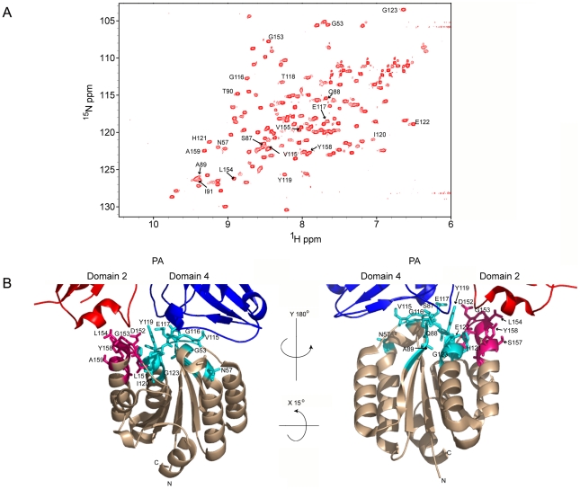
Figure 1. A) 1H-15N TROSY-HSQC of the ANTXR2 VWA domain.
Cross-peaks with labeled assignments represent receptor residues at the PA domain 2 and domain 4 interaction surfaces. B) The crystal structure of the interface between monomeric PA83 bound to ANTXR2 (PDB 1T6B; Protein Data Bank), showing the base regions of domain 2 and 4 of PA. Representative PA contact residues of ANTXR2 are indicated: domain 2 contact sites are in hot pink and domain 4 contacts are in cyan. All images were generated using PymolX11 (DeLano Scientific, San Carlos, CA).

