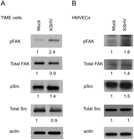
Figure 2. FAK and Src are activated during KSHV latency in endothelial cells.
TIME cells (A) or primary dHMVECs (B) were mock- or KSHV-infected. At 48 hpi, cell extracts were analyzed by immunoblot analysis with the indicated phospho-specific or total-protein antibodies. Blots were stripped and probed with an antibody to β-actin as a loading control. The numbers below the lanes indicate the relative abundance of each major band. These experiments were repeated three times with similar results.

