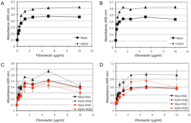
Figure 4. KSHV latent infection promotes adhesion to fibronectin and vitronectin in an integrin-dependent fashion.
TIME cells were mock- or KSHV-infected and 48 hours post-infection cells were plated onto 96-well plates coated with fibronectin (A) or vitronectin (B). Cells were allowed to adhere for 90 minutes after which non-adherent cells were washed off. The number of adherent cells was determined by alkaline phosphatase activity. Mock-infected cell adhesion is indicated with squares and KSHV-infected cell adhesion is indicated with triangles. C and D) 48 hours post-infection, mock- (squares) and KSHV- (triangles) infected cells were pretreated with RGD-containing (red lines) or control peptide (RAD for (C) and RGE for (D), black lines) for 15 minutes on ice before assaying for adhesion to fibronectin (C) or vitronectin (D).

