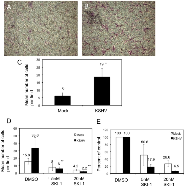
Figure 5. KSHV latent infection promotes endothelial cell transwell migration in a Src-dependent fashion.
Cells were infected with mock or KSHV and, 48 hours post-infection, were plated on fibronectin-coated transwells. Cells were allowed to migrate through the pores at 37°C for 1.5 hours and subsequently fixed and stained with crystal violet. The cells on the upper side of the membrane were removed with a cotton swab, the membranes were mounted on slides and the cells that migrated were photographed. A and B) Representative images of migrated mock- (A) and KSHV-infected (B) cells. C) Quantification of a representative experiment showing the number of migrated cells per field for 10 fields counted per transwell. Asterisk indicates a p-value of less than 0.05 compared to mock-infected cells. D–E) Cells (48 hpi) were treated with the Src kinase inhibitor SKI-1 at the time of plating on fibronectin-coated transwells and are presented as number of cells per field (D) and percent inhibition by SKI-1 (E). Double asterisks indicate a p-value of less than 0.005 compared to DMSO control.

