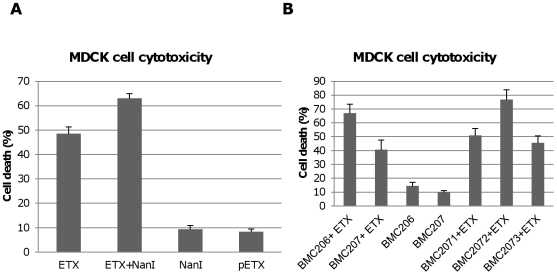
Figure 7. Sialidases increase cytotoxicity for MDCK cells.
Panel A, percent of dead MDCK cells after treatment at 37°C for 1 h with 5 µg/ml ETX, 5 µg/ml pETX, 0.005 U/ml NanI sialidase or a mixture of 5 µg/ml ETX and 0.005 U/ml NanI sialidase, Panel B, percent of dead MDCK cells after the cells were treated for 1 h at 37°C with 5 µg/ml of ETX dissolved in BMC206, BMC207, BMC2071, BMC2072 or BMC2073 culture supernatants. BMC206 and BMC207 culture supernatants without ETX supplementation are shown for comparison. The results shown represent the average of three repetitions; the error bars indicate standard errors of the means.

