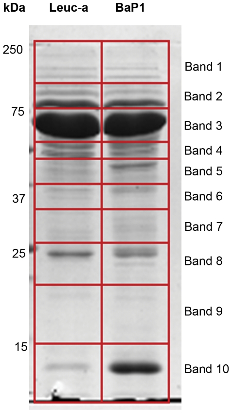
Figure 7. SDS-PAGE of exudates samples collected from mice injected with either BaP1 or leuc-a.
Samples corresponding to 20 µg protein of exudates collected 15 min after injection of SVMPs were electrophoresed on a 4–20% gradient gel followed by staining with Coomassie Blue. Molecular mass markers are depicted to the left. Gel lanes were cut into ten equal size slices for further proteomic analysis (see Methods for details).

