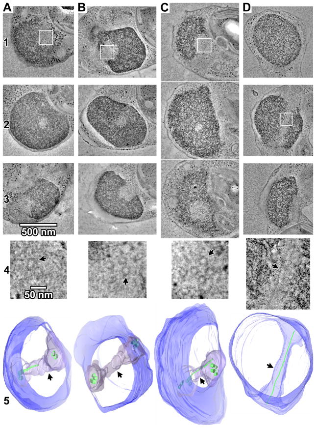
Figure 1. O. tauri has only a few spindle microtubules.
(A–D) Tomographic slices (19 nm thick) through merged serial-section tomograms of MG132-treated, high-pressure frozen, freeze-substituted O. tauri cells. The entire nuclei and spindles are reconstructed in the merged tomogram. The cells in columns A-C were sectioned nearly transverse to the spindle tunnel, while the cell in column D nearly longitudinal. Rows 1 –3 show tomographic slices through upper, middle, and lower plastic sections of the nucleus. Row 4 shows enlargements of the areas boxed in white in Rows 1 or 2, with small rotations out of plane to enhance the visibility of the spindle MTs. Arrows in row 4 indicate example MTs. Row 5 shows 3-D segmentations of the chromatin boundary (blue), spindle tunnel (arrowheads), and MTs (green). Note that the tunnel-spanning portion of long MTs have incomplete walls and are modeled as thin rods instead of tubes. The cell in (B) is shown in greater detail in Movie S1.

