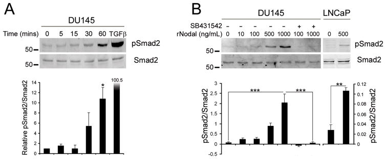
Figure 3. Nodal Signaling in Prostate Cancer Cell Lines.
(A) DU145 cells were treated for 5–60 mins with 1 μg/mL recombinant Nodal or 30 min with 1 ng/mL TGFβ1. Western blots of pSmad2 and total Smad2 levels were quantified using densitometry. Nodal significantly increased Smad2 phosphorylation at 60 mins (One Way Anova with Tukey’s posthoc analysis, n=2, *P<0.05). (B) DU145 cells were treated for 60 min with 10–1000 ng/mL Nodal and 10 μM SB431542 (+) or DMSO control (−). LNCaP cells were treated with 500 ng/mL Nodal for 6 hrs with fresh Nodal added every 2 hrs. There was a significant increase in pSmad2 with 1 μg/mL Nodal in DU145 cells and significant decrease when SB431542 was added (One Way Anova with Tukey’s posthoc analysis, n=3, ***P<0.001). There was also a significant increase in Smad2 phosphorylation in LNCaP cells treated with Nodal (t test, n=3, **P<0.01).

