
Figure 4.
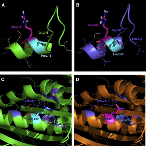
3D Structure of KIF22 and the Effect of Mutations
(A) The wild-type structure was obtained by running MD simulation.
(B) The mutant structure with Pro148Leu mutation from MD simulation revealed two new hydrogen bonds; the hydrogen bond between NH of Arg148 and C = O of Gly145, and the hydrogen bond between NH of Leu148 and C = O of Leu138.
(C) Arg149 forms a hydrogen bond with Gln101 and Tyr104 residues in an α-helix located near ATP binding site and also with loop residue Pro144.
(D) The p.Arg149Gln mutation would disrupt the hydrogen bonds with the α-helix of Gln101 and Tyr104. The red dotted line symbolizes a hydrogen bond.