
Figure 1.
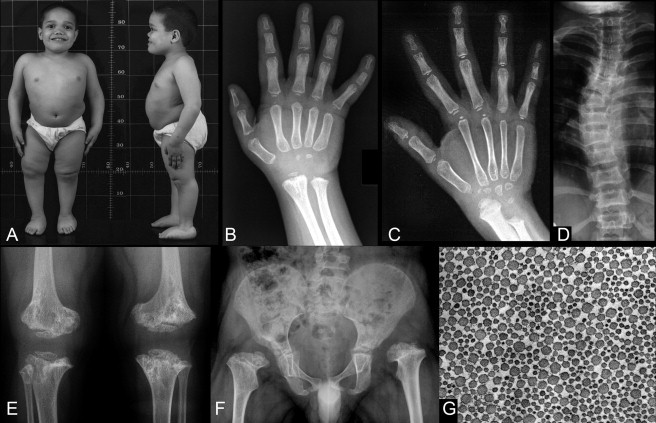
Morphologic Features of Lepto-SEMDJL
(A, B, D, E and F) are all from subject 2 (family 1) at age 7.
(A) In this boy, stature is markedly below the normal range, with short-trunk type disproportion. There is frontal bossing with flattening of the face and a sunken nasal bridge. There is left hip subluxation (F), leg length difference, and right genu varum (A). Joint laxity is indicated by the scoliosis and the flat feet.
(B) The hand radiographs of this boy; there is a very marked delay in the maturation of all epiphyseal centers and of the carpal bones, as well as metaphyseal irregularities at the distal radius and ulna.
(C) The hand X-ray of an unrelated boy, age 10. Also in this individual, there is a marked delay in all secondary ossification centers and there is shortening of the distal ulna. The proximal phalanges and the metacarpals are slender; this feature, leptodactyly, that becomes apparent only over time, is characteristic for this bone dysplasia.
(D) The moderate platyspondyly and the scoliosis (ligamentous laxity).
(E) The marked dysplasia of the metaphyses at the knee (distal femur, proximal tibia) and at the same time the small and dysplastic epiphyses.
(F) A similar pattern at the proximal femurs with shortening of the femoral necks and the presence of epiphyses that are barely visible and markedly small for age. The acetabula are not well developed; they are less well developed on the left than on the right; the left hip is subluxated because of the acetabular dysplasia and associated ligamentous laxity.
(G) An electron microscopy image of a tendon biopsy section (subject 6 in Table 1) at right angle to the collagen fiber (magnification, approximately 5000×). The diameter of the fibers shows a significant variability.