
Figure 1.
Clinical Description of the Affected Family Members
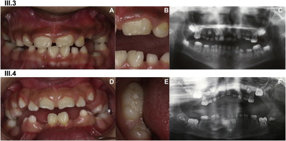
A clinical description of individuals III.3 (A, B, and C) and III.4 (D, E, and F) shows major dental developmental abnormalities in tooth number, size, shape, structure, eruption, and resorption, as seen in the intraoral photographs (A, B, D, and E) and the panoramic radiographs (C and F).
(A and B) (A) shows an intraoral view of III.3 (10 years old). Beside the microdont primary and permanent teeth, which show spaced dentition, double tooth formation (notched and macrodont) is visible on permanent central incisors 21 and 31; 21 shows a vestibular abnormal relief. These anomalies are clearly seen on (B) in an enlargement of the left incisor region.
(C) A panoramic radiograph shows III.3, who is missing the following permanent teeth: 18, 15, 24, 25, 28, 48, 45, 44, 43, 32, 33, 34, 35, and 38. The primary and permanent molars are taurodont. The roots are extremely short and are slightly more developed in the permanent dentition but are, however, conical with sharp endings. The pulp has a flame-like shape. The enamel is very thin and has limited contrast compared to the dentin in the X-Ray. Teeth 64, 65, 74, and 75 are reincluded.
(D) Intraoral view of III.4 (9.5 years old). Double tooth formation (notched and macrodont) is visible on the permanent central-upper-left incisor (21). The lower arch seems interrupted in the area of missing teeth (45, 43, 42, 32, 33, 34, and 35). Teeth 85 and 75 are reincluded, indicating ankylosis in the alveolar bone.
(E) A close-up on macrodont tooth 46 (lower-right permanent first molar) shows extra cusps and an elongated crown on its mesiodistal axis.
(F) A panoramic radiograph of III.4 at 5 years old shows oligodontia—13 permanent teeth are missing (18, 15, 25, 28, 48, 45, 43, 42, 32, 33, 34, 35, and 38)—and extreme microdontia of all the primary teeth. Note the short and sharp roots and the hypodeveloped alveolar bone.