
Figure 2.
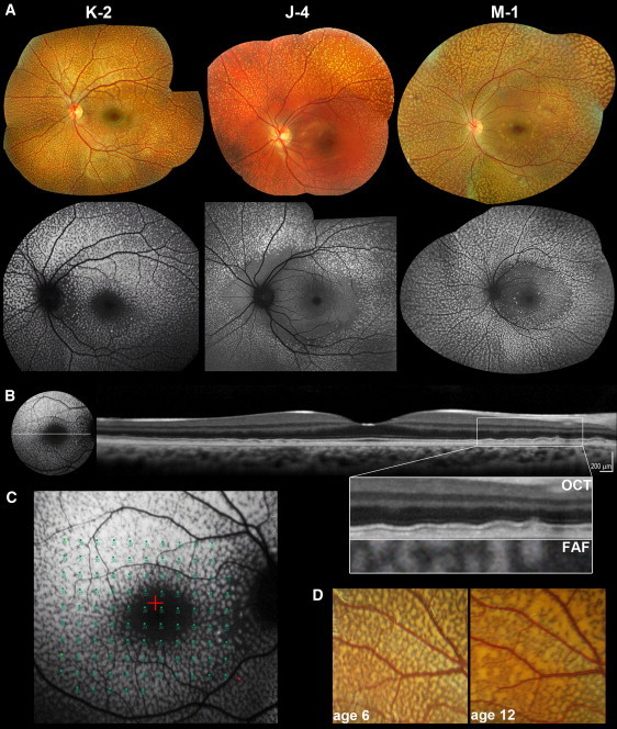
Retinal Imaging of Individuals with Benign Fleck Retina and PLA2G5 Mutations
(A) Color photographs and corresponding fundus autofluorescence (FAF) images of the left fundi of subjects K-2 (aged 12), J-4 (aged 12), and M-1 (aged 39). On fundus photography, multiple yellow-white flecks of various sizes are observed. FAF reveals hyperautofluorecent lesions corresponding in location with the flecks. The macula is relatively spared in subjects J-4 and M-1 but not in K-2, in whom only the fovea appears to be unaffected. This might reflect a more detrimental effect of the c.185G>A (p.Trp62X) mutation in the homozygous state (subject K-2) as opposed to homozygous missense (c.133G>T [p.Gly45Cys] in subject J-4) or compound heterozygous (p.Gly45Cys and c.383delA, p.Gln128ArgfsX88 in M-1) mutations.
(B) FAF imaging and linear spectral domain optical coherence tomography (OCT) scan of the left retina of subject K-2. Deep, discrete, hyper-reflective deposits, more obvious at the edge of the foveal scan, are observed. The panel with an enlarged image of the boxed region shows the outer retina and RPE in detail. The lesions are located posterior to the hyperreflective band corresponding to the photoreceptor inner/outer segment junction and do not disrupt it. An overlay of OCT with FAF is also presented. Deposits are spatially associated with the hyperautofluorescent lesions and thus correspond to the flecks.
(C) Functional assessment of the central retina in subject K-2. Static-perimetry testing (threshold sensitivities from 0 to 20 dB, test spot size Goldmann III) results overlaid with FAF are presented. Retinal sensitivity was normal.
(D) Longitudinal data showing evolution of fleck-like lesions over time. Magnified view of fundus photographs from the left eye (vascular arcades) of subject K-2 at ages 6 and 12. Flecks increase in number and size and become more confluent.