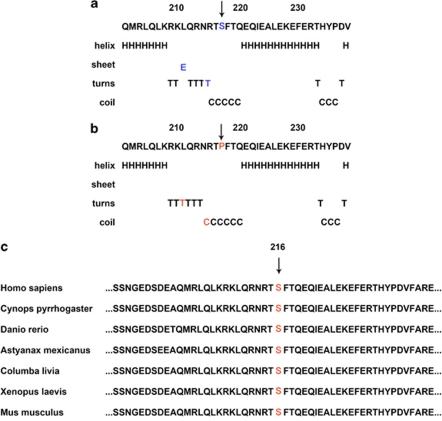
Figure 5.
The effect of p.S216P on the secondary structure of the PAX6 using the GOR method and sequence alignment portion of the HD spanning the p.S216 of the PAX6 of human with other species. (a) The secondary structure of wild-type PAX6 around the site S216. (in blue). (b) The secondary structure of mutant P216 of the PAX6 of the corresponding region (in red). (c) Sequence alignment portion of the HD spanning the p.S216 of the PAX6 of human with other species. The mutation site is boxed in red letters. The color reproduction of this figure is available at the Eye journal online.

