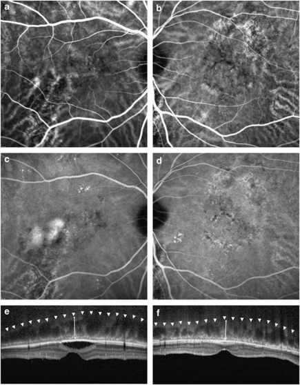
Figure 1.
Representative ICGA and EDI-OCT of a patient with unilaterally active CSC. Choroidal vascular dilation and hyperpermeability on angiography and increased choroidal thickness on OCT were noted in the unaffected fellow eye (b, d, f), as well as in the eye with active CSC (a, c, e). Arrowheads indicate the inner sclera border and arrows demonstrate the estimated choroidal thickness from the outer border of the Bruch membrane to the inner scleral border.

